What is fluoroscopy? X-Rays, 'ALARA' & more
The term 'fluoroscopy' may sound intimidating, but also known as the 'X-ray' procedure, it is amongst the most well-known radiological procedures in the world today.

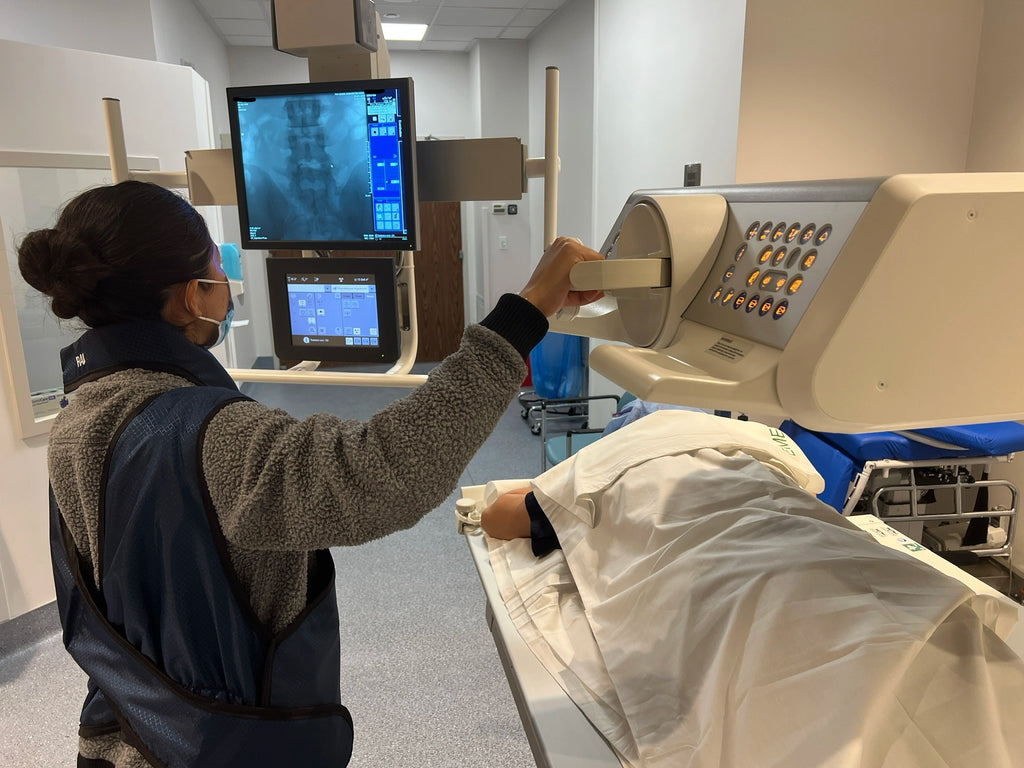
A radiologist performs a fluoroscopy procedure on a patient to see the internal workings of their pelvic region in real-time.
What is fluoroscopy?
A fluoroscopy procedure is a type of medical test that uses X-rays to create real-time images of your body's internal structures. It provides a continuous stream of x-ray images that creates a mini-movie showing what is happening inside the body for a snapshot of time.
What can be diagnosed with fluoroscopy?
A radiologist would use fluoroscopy to visualize and evaluate the function and structure of organs, tissues, and bones in real-time. Minimally-invasive procedures can also be accomplished using fluoroscopy.
Here are some examples of how a radiologist might use fluoroscopy:
Swallowing and upper gastrointestinal procedures:
A radiologist might use fluoroscopy to perform a barium swallow, which involves swallowing a liquid containing barium sulfate, a contrast agent. As the patient swallows, the radiologist can visualize the barium moving through the esophagus and into the stomach to diagnose conditions such as gastroesophageal reflux disease (GERD), hiatal hernia, ulcer disease, or tumor. The small intestine can be studied as well.
Lower gastrointestinal procedures
A radiologist might use fluoroscopy to perform procedures such as an enema which evaluates the large intestine for abnormalities such as tumors or blockages.
Angiography
A radiologist might use fluoroscopy to inject a contrast agent into an artery or vein to visualize blood flow. The radiologist can then evaluate the blood vessels for abnormalities such as aneurysms or blockages.
Orthopedic procedures
A radiologist might use fluoroscopy to guide orthopedic procedures such as joint injections or biopsy with real-time accuracy.
Spinal procedures
A radiologist can use fluoroscopy to guide lumbar puncture or myelogram, which can be used for diagnosis of central nervous system infection, tumors, or degeneration.
Other advanced interventional procedures
An interventional radiologist utilizes fluoroscopy to access and treat nearly any organ system from head to toe.
What can I expect on the day of my procedure?
When you arrive for the procedure, you'll be asked to change into a hospital gown and remove any jewelry or metal objects that may interfere with the imaging. You'll then be taken to a special room where the radiologist will perform the procedure.
During the fluoroscopy, you'll lie on a table and a special camera called a fluoroscope will be positioned over the area of your body that needs to be imaged. The radiologist will then use X-rays to create real-time images of your body as you move or as the radiologist manipulates the fluoroscope.

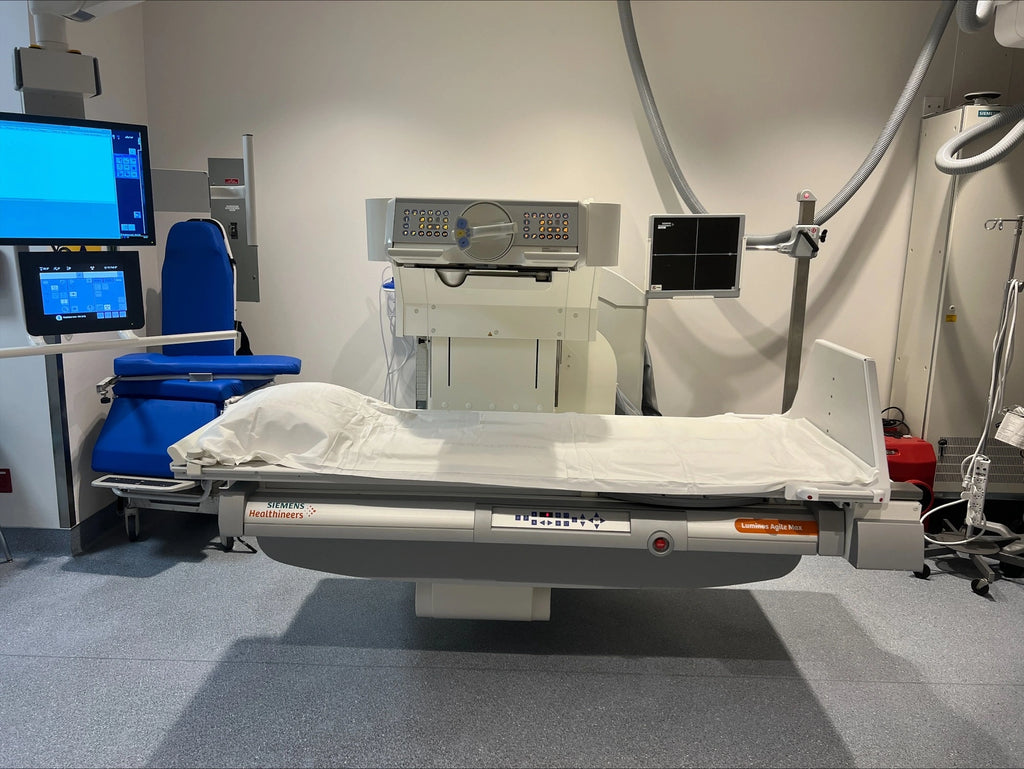
The X-ray table may not look like much, but it is a powerful tool that is integral to making accurate medical diagnoses worldwide!
You may be given a contrast agent, which is a special dye that helps certain structures or organs show up more clearly on the X-ray images. The contrast may consist of barium sulfate or iodine solution and be given orally, intravenously, or via an enema–depending on the area being examined!
Of note, it's important to hold still during the procedure to ensure clear images are obtained! You may be asked to hold your breath for a few seconds at a time to reduce movement and obtain clear images.
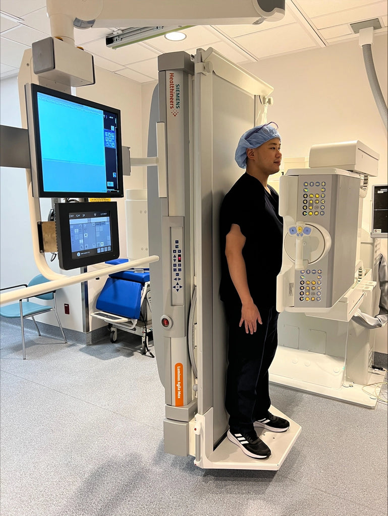
You may also be asked or helped to change position on the exam table, stand or to lie down. This is completely normal.

The procedure usually lasts only a few minutes, but the exact duration will depend on the area being imaged.
After the procedure, you may be asked to wait for a short time while the radiologist reviews the images.
The radiologist will then provide the results of the procedure to your healthcare provider, who will then share them with you!
Is fluoroscopy safe?
While radiation exposure from a single fluoroscopy procedure is generally considered safe, repeated exposure over time can increase the risk of cancer and other radiation-related health problems. It is important for doctors and other healthcare professionals to carefully evaluate the risks and benefits of any medical imaging test before recommending it to a patient, and to use the lowest possible dose of radiation necessary to obtain the needed information.
This is the 'ALARA principle', which is a radiation safety principle that stands for "As Low As Reasonably Achievable." It is a fundamental principle in radiation safety that radiation exposure is not completely avoidable, but steps can be taken to minimize exposure to patients and healthcare workers.
The ALARA principle is based on three key concepts:
-
Justification: The use of radiation should be justified by weighing the benefits of the procedure against the potential risks.
-
Optimization: Radiation doses should be optimized to achieve the desired diagnostic or therapeutic result while minimizing radiation exposure.
-
Dose limits: Radiation doses should be kept within regulatory dose limits to ensure that exposure remains as low as reasonably achievable.
Radiologists at LAIIC practice ALARA to maximize your safety while ensuring the best possible outcome for your diagnostic procedure and treatment.
In conclusion
Having a fluoroscopy procedure scheduled to diagnose a health condition is very, very common. In fact, According to the Pan American Health Organization, there are an estimated 3.6 billion diagnostic X-ray exams performed annually.
That makes the x-ray the most common diagnostic imaging exam performed worldwide!
If you have any questions, or would like to schedule a radiology appointment in Southern California, please give us a call or schedule an appointment with us online!

